[dkpdf-columns columns=”3″ equal-columns=”false” gap=”10″]
Introduction
Metabolic Syndrome (MetS), also known as insulin resistance syndrome or syndrome X, is defined as a combination of abnormalities resulting as a consequence of insulin resistance and abnormal adipose tissue regulation. Its features include obesity, high blood pressure, elevated blood sugar levels and high triglycerides. The overall clinical manifestation of the condition is an increase in the risk of cardiovascular disease, type-2 diabetes, non-alcoholic fatty liver disease (NAFLD) and obesity-related cancers.1
Insulin resistance results in defective insulin receptor signalling and impaired gluconeogenesis in the liver, fat and muscles cells. Adipose tissue dysregulation leads to an increase in free fatty acids and accumulation of (intra-abdominal) visceral fat which promotes inflammation due to the excessive release of pro-inflammatory cytokines.
Independent of sex and ethnicity, the likelihood of developing metabolic syndrome is greater with increasing BMI. The occurrence of this condition is, however, not restricted to obese individuals of either sex. The concept of metabolic syndrome in non-obese individuals refers specifically to individuals with a BMI of 19-29.9 Kg/m2 who display characteristics of metabolic syndrome such as insulin resistance, dyslipidaemia and visceral adiposity. In a 2017 study of 1168 Malaysian teachers, Lee S.C et al. (2017) reported an 8.3% prevalence of metabolic syndrome occurring among non-obese participants.3
Prevalence
The prevalence of metabolic syndrome is increasing in line with the rising level of obesity worldwide.Data from the National Health and Nutrition Examination Survey (NHANES) database in 2002 revealed a prevalence of 34.5%.4 The Framingham Heart study recorded a prevalence of 26.8% in men and 16.6%in women. Although the prevalence of MetS is higher in developed nations due to the higher prevalence of obesity, the condition is recognised as a global problem with rising incidence in Asia, Africa and Latin America.5
Pathogenesis
Obesity, especially visceral adiposity is a chronic low-grade inflammatory condition. Unlike subcutaneous fat, visceral (intra-abdominal) fat strongly correlates with inflammation, dysfunctional adipose tissue regulation and secretion of pro-inflammatory cytokines, typically associated with the occurrence of metabolic syndrome.
Hypertrophied dysregulated adipocytes secrete pro-inflammatory cytokines resulting in oxidative stress and metabolic derangements such as hyperglycaemia, dyslipidaemia, endothelial dysfunction and atherogenesis. Metabolic abnormalities associated with MetS can be detected (and monitored) by the presence of serum biomarkers. Detection of these early biomarkers could be of use in the management of diabetes and metabolic syndrome in high-risk populations.6
Biomarkers for Metabolic syndrome
Leptin – Discovered in 1994, leptin is a hormone produced predominantly by adipose cells. Leptin
is responsible for energy expenditure and the regulation of appetite (hunger inhibition). Its signal from energy (adipose) stores within the body exerts a negative-feedback effect on appetite and energy intake through receptors in the arcuate nucleus of the hypothalamus.
Obese individuals, however, develop leptin resistance (akin to insulin resistance in type-2 diabetes patients) despite high circulating levels of leptin, resulting in further weight gain due to a loss of hunger inhibition. Elevated leptin is therefore associated with obesity and insulin resistance.The level of this biomarker is proportional to the mass of adipose cells present and therefore predictive of obesity-related chronic conditions such as metabolic syndrome.7
Adiponectin – Unlike Leptin, adiponectin is produced only by adipocytes. It promotes insulin sensitivity and lipid oxidation and protects the endothelial cells against atherosclerotic vascular damage. Low plasma levels of adiponectin are observed in metabolic syndrome and individuals with visceral adiposity, hypertension and type 2 diabetes.8
Leptin/Adiponectin Ratio (LAR) – High LAR is a more sensitive biomarker for metabolic syndrome than either Leptin or Adiponectin in isolation. LAR is directly associated with MetS and positively correlates with all the individual components of MetS.9
Uric acid – Uric acid is a marker for oxidative stress. It demonstrates pro-inflammatory and pro-oxidant activity, resulting in tissue damage. Hyperuricemia is a risk factor for atherosclerosis,hypertension and dyslipidaemia. The level of uric acid is elevated in MetS and also in all the individual components of MetS.10
Diagnosis
Although the concept of metabolic syndrome provides a practical and useful way of identifying patients with multiple risk factors for developing diabetes and other forms of cardiovascular disease,MetS is often not diagnosed in clinical practice as an individual disease entity.11 Instead, the cardiovascular and other standard components of the syndrome are usually identified and treated as separate disease conditions. Not often mentioned is the high degree of prevalence of conditions such as hypogonadism, also observed in obese individuals with metabolic syndrome.12 Recognising MetS as a disease cluster is essential, to ensure early intervention and prevention of
long-term complications and morbidity.13
Several diagnostic criteria have been proposed for the definition and identification of metabolic syndrome with input from – the National Cholesterol Education Program Adult Treatment Panel III NCEP ATPIII), the International Diabetes Federation (IDF) and the World Health Organisation (WHO).More recently, the National Heart, Lung and Blood Institute (NHLBI) and the American Heart Association (AHA) proposed a revised set of ATPIII guidelines recommending the diagnosis of metabolic syndrome based on 3 out of 5 criteria listed in table 1:
Patient X’s presentation with erectile dysfunction (suggestive of hypogonadism) is however
CASE STUDY 1
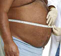
Patient X – A 38-year-old hypertensive gentleman (on ACE-inhibitor treatment) referred by his GP, to the endocrine clinic with a history of erectile dysfunction. He drinks several units of alcohol a day.
On clinical examination (fig 1), he was morbidly obese with a BMI 42kg/m2. His blood pressure was 165/90mmHg. He had features typical of insulin resistance – prominent supraclavicular fat pad, acanthosis nigricans and skin tags on his neck. Biochemical investigation revealed the following results – total testosterone level – 2.0 nmol/l (8.4-27.4), cholesterol – 7.2 mmol/l (0-5), triglyceride – 10.2 mmol/l (0.3-1.7), ALT – 102 U/L (9-55), ALP – 200 U/L (30-130) plasma glucose – 8.9 mmol/l and HBA1c – 7.8% (63 mmol/mol). Liver ultrasound scan and abdominal MRI revealed fatty liver disease and extensive visceral adiposity.
Diagnosis – This is a classic case in which the patient fulfils the ATPIII criteria for diagnosing metabolic syndrome. Further investigationconfirmed diagnoses of dyslipidaemia, type-2 diabetes, and non-alcoholic fatty liver disease (NAFLD).
Treatment – An aggressive change in lifestyle was recommended – smoking and alcohol cessation, dietary changes and regular exercise,in combination with GLP-1 analogue therapy (for weight loss).14 The patient lost 26kg in weight (BMI reduction to 34). 12 months later,his medication was discontinued. His blood pressure was 125/70, HBA1c – 5.6% (38mmol/mol), serum cholesterol – 4.2mmol/l,Triglyceride levels – 1.4mmol/l, testosterone – 9.2nmol/l and liver function tests (LFT) returned to normal levels.
Table 2: Metabolic syndrome, Obesity and Hypogonadism
atypical in the present context.15 Table 4 outlines the essential relationship between hypogonadism and obesity in metabolic syndrome.
Clinical implications
Both cases (outlined in tables 2 and 3) describe individual scenarios of patients with a characteristic
diagnosis of metabolic syndrome but presenting in different ways. Both patients have high triglyceride levels, type 2 diabetes, hypertension, and non-alcohol fatty liver disease and are therefore at significantly increased risk of cardiovascular morbidity.
CASE STUDY 2
Patient Y– A 67-year-old female was referred to the diabetes clinic with a recent diagnosis of type-2 diabetes, for which she has already been prescribed 1g Metformin twice daily and Canagliflozin 300mg once a day. She has a background history of hypertension and hypercholesterolemia. Her current medication includes Ramipril 5mg daily, Atorvastatin 20mg daily and Aspirin 75mg.On clinical examination (fig 2), BMI was 25.2kg/m2 and BP 155/90mmHg. She has features suggestive of peripheral
On clinical examination (fig 2), BMI was 25.2kg/m2 and BP 155/90mmHg. She has features suggestive of peripheral neuropathy and peripheral vascular disease. Monofilament and vibration tests were negative in the distal regions. Dorsalis pedis and posterior tibial pulses were absent on (hand-held) Doppler examination. Blood biochemistry showed the following – HBA1c – 9.2%, plasma glucose – 10.5 mmol/l, cholesterol – 6.5mmol/l, triglyceride –6.2mmol/l, ALP – 190U/L and ALT – 99U/L. Liver ultrasonography revealed features of fatty liver.
Diagnosis – Type-2 diabetes (newly diagnosed/inadequately controlled), peripheral vascular disease, coronary artery disease,hypertension, dyslipidaemia and non-alcoholic fatty liver disease (NAFLD).
Treatment – Patient Y was started on insulin therapy (Novomix 30), The dose of Atorvastatin was increased to 40mg daily (a fibrate was subsequently added to the regimen), Ramipril was increased to 10mg daily and Amlodipine was added to achieve improved blood pressure control. Further investigations revealed occluded coronary and popliteal arteries. She required angioplasty to both occluded arteries.
Table 3: Metabolic syndrome in the non-obese patient
CPD UPDATEM
The origin of hypogonadism in MetS is due mainly to an increase in aromatase activity that arises from the excessive accumulation of fatty tissue in obese individuals.16
ypogonadism in the metabolic syndrome is mediated by Aromatase, an enzyme produced in fat (adipose) cells and associated with increased conversion of testosterone to oestradiol (testosterone-oestradiol shunt). The decreased levels of bioavailable testosterone (and a corresponding rise in oestradiol) encourages the preferential deposition of further abdominal fat,leading to a further reduction of circulating testosterone and progressive hypogonadal state. A loss of compensatory (feedback) increase in gonadotrophin production in MetS (due to inhibition of the hypothalamo-pituitary axis) often results in a progressive cycle of hypogonadotropic hypogonadism.17
Aggressive lifestyle intervention achieved successful treatment outcomes (and risk reduction) in Case 1. Despite the lack of symptoms in Case 2,Patient Y had already developed coronary artery disease and peripheral atherosclerosis.
The second case illustrates the concept of metabolic syndrome in a non-obese individual. Despite a normal BMI, she fulfils the ATP III criteria for the diagnosis of metabolic syndrome. As previously discussed,metabolic syndrome may occur in both obese and non-obese individuals.
The most appropriate treatment approach in metabolic syndrome is to screen the individual for all constituent elements of the MetS cluster of diseases, initiate aggressive lifestyle modification and specific treatments
where necessary, and maintain an overall objective of reducing cardiovascular morbidity and mortality.
A detailed history and clinical examination should always be
Recognising metabolic syndrome as a disease cluster is essential to early intervention and prevention of long-term morbidity
performed to detect the presence of type 2 diabetes, hypertension,dyslipidaemia, fatty liver disease,obstructive sleep apnoea, depression and hypogonadism.
Assessment of 10-year cardiovascular risk using a valid risk assessment score such as QRISK 2 or Framingham Risk score is essential to target individuals at increased risk of developing cardiovascular disease.
Treatment
The three components of treatment are as follows – (1) Aggressive lifestyle modification and medical therapy for established risks, (2) management of obesity and hyperglycaemia; and (3) regular screening for cardiovascular disease.18

Diet and Lifestyle: Healthy diets with a caloric restriction such as Mediterranean diet, low fat and low glycaemic index food; smoking cessation; reduction in alcohol intake and increased physical activities are
the recommendations to prevent metabolic syndrome and improve cardiovascular health.19
Medical therapy: Statins and fibrates to treat dyslipidaemia, ACE inhibitors, a calcium antagonist and β blockers to treat hypertension;Metformin, GLP-1 agonist and SGLT2 inhibitors to treat obese MetS patient with diabetes; bariatric surgery for morbidly obese patients and antiplatelet therapy for patients at high risk of developing ischaemic heart disease.
Screening for cardiovascular disease: Patients with symptoms should be assessed with an ECG, echocardiogram and coronary angiogram to help detect the presence of established coronary artery disease and left ventricular systolic dysfunction.
Summary
Metabolic syndrome is a cluster of abnormalities occurring as a consequence of defective insulin signalling, abnormal adipose tissue regulation and chronic low- grade tissue inflammation leading to oxidative stress. The primary mediator of this condition is insulin resistance. MetS is a significant risk factor for cardiovascular disease and type 2 diabetes.
The main clinical features of metabolic syndrome are abdominal obesity, hypertension, dyslipidaemia, hyperuricaemia and hyperinsulinaemia. The National Heart, Lung and Blood Institute (NHLBI) and the American Heart Association (AHA) proposed revised ATPIII guidelines on the basis of 3 out of 5 criteria (table 3).
The correct management approach in patients with metabolic syndrome is based on aggressive lifestyle intervention and treatment to prevent the development of type-2 diabetes and associated cardiovascular risk. In those with established risks, targeting the individual components of MetS with medical therapy is essential.
[/dkpdf-columns]
read moreDisclosure forms provided by the author are available at NEJM.org.
Editor’s note:
Author Affiliations
Supplementary Material
| Disclosure Forms | 83KB |
Add your Comment


Add your Comment
Leave a Reply
You must be logged in to post a comment.
BHQJ 2018 ; 001:34-36
Related Article 
Medical Negligence & the Law5th May 2018 . Atrogenic harm is a matter of significant concern in Nigeria admin
Colorectal Cancer Overview5th May 2018 . [dkpdf-columns columns="3" equal-columns="false" gap="10"] Introduction Colorectal cancer is a major admin
Funding Healthcare Services in Nigeria – A conundrum of demand, policy and supply!5th May 2018 . [dkpdf-columns columns="3" equal-columns="false" gap="10"] Doctor, I happy say na you admin
5 “Provocations” of Healthcare Quality Reform6th May 2018 . [dkpdf-columns columns="3" equal-columns="false" gap="10"] n the four decades since he admin
Health Insurance, Activism & Urgent Change6th May 2018 . [dkpdf-columns columns="3" equal-columns="false" gap="10"] AR: Dr Soyinka, it’s wonderful to admin
Anne Olowu talks about her “Masterclass” experience6th May 2018 . [dkpdf-columns columns="3" equal-columns="false" gap="10"] As I suspect is the case admin
Stomach & Oesophageal Cancer in Nigeria6th May 2018 . [dkpdf-columns columns="3" equal-columns="false" gap="10"] Gastric and Oesophageal (Upper GI) cancers admin
Setting out the Stall!7th May 2018 . [dkpdf-columns columns="3" equal-columns="false" gap="10"] "The drawbacks of our false knowledge admin
Leave a Reply
You must be logged in to post a comment.