[dkpdf-columns columns=”3″ equal-columns=”false” gap=”10″]

Gastric and Oesophageal (Upper GI) cancers remain a major health challenge worldwide.Combined, these cancers are
responsible for approximately 1,407,378 (10%) cancer deaths per annum.Nigeria has a reported 101.3 cancer cases per
100,000 (UK 272.9 cancers per 100,000).Despite such reported low cancer rates, we know that due to its large population size and low levels of socioeconomic development,Nigeria and other low and middle-income countries in sub-Saharan Africa and beyond, together carry a substantial and disproportionate burden of cancer, all in all
accounting for over half (57%) of the world’ stotal.
In the 21st century, the diagnosis and management of Upper GI cancers is a multidisciplinary task, promoting accurate diagnosis and appropriate staging of disease. Treatment modalities typically involve a combination of
chemotherapy, radiotherapy and surgery. The challenge, however, remains detection of the disease at an early stage.
It is a well-known fact that by the time symptoms are reported in the majority of patients, Upper GI cancers are usually at an advanced stage and not infrequently metastasised incurably to distant organs like the liver and lungs. The negative impact of such spread on outcomes observed in this scenario is remarkably profound.
Regions of the world with a high incidence of such cancers, like Japan and China, have invested considerably in targeted screening programs aimed entirely at early detection usually at the asymptomatic, initial period of the
disease, during which patients are usually without symptoms.1
The incidence of gastric and oesophageal cancer in Nigeria remains unclear. There have been retrospective studies reporting this incidence to be low. However, as with other forms of cancer in this part of the world, a lack of cancer reporting registries, major deficiencies in access to adequate endoscopy, the absence of high-quality histopathological services and low levels of community awareness, means we do not really know the true situation regarding
Case Study 1
It was a weekday morning on the day that I received a call from the hospital manager about a 42-year-old man who had called up just a few moments before. He was complaining of a single episode of transient dysphagia (difficulty swallowing) while having dinner the evening before.
During a telephone consultation, I was able to understand that his symptom was accompanied by some transient pain at the time. He recalls drinking several mouthfuls of water appeared to aid in its rapid resolution and left him feeling as he described it with ‘a wound-like feeling’ in his upper abdomen (epigastric region) whenever he attempted to swallow after the episode was over. He admitted that he was yet to try anything solid since the event of the evening before but was remarked that he was tolerating fluids ‘OK’. There was no prior history of anything significant aside from a history of infrequent reflux disease.My immediate thoughts went towards a possible hiatus hernia as the likely underlying problem. I recommended a clinic review,possibly an early endoscopy along with acid suppression therapy.
Later that day, he came to see me in clinic. He was a fit and well-looking gentleman, currently working as a high-ranking executive of an international organization There was nothing further in the history of note. He was in full agreement with the plan for an endoscopy and we proceeded later that day to do the needful.
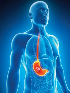
To my shock and surprise, I discovered a 1cm malignant ulcer in the cardia of the stomach, just beyond the gastro-oesophageal junction. I placed him on high dose acid suppression and a soft diet, pending pathology results. The histopathological analysis returned and revealed moderately differentiated adenocarcinoma of local origin. Staging CT scan of the chest, abdomen and pelvis showed only mild thickening at the cardia. The upper GI MDT case review agreed that a decision to proceed straight to surgery was an appropriate treatment option.
Consent was given for the procedure to proceed and we performed a laparoscopically-assisted vagus nerve-sparing proximal gastrectomy and Merendino Reconstruction (Interpositional Jejunum–oesophagojej and Gastro-Jej anastomosis)4. He made an uneventful post-surgical recovery, leaving the hospital 4 days later on a liquid diet for 2 weeks. The postoperative histology revealed a yPT1N1Mx adenocarcinoma of the stomach with 1 out of 15 LN involved and a focus of microvascular invasion. The Upper GI MDT recommended 4 cycles of Adjuvant FLOT chemotherapy. He is currently going through this and appears to be coping well.
Case Study 2
A 44-year old gentleman was referred with a 10-month history of persistent epigastric pain, worsening reflux disease, easy satiety and weight loss. He had undergone an endoscopy in a different centre 2 months previously and was apparently advised he may have ‘Menetrier’s disease’ of the stomach. Biopsies, at the time of his consultation with me, were not available.
He had no prior or family history of significance.Clinical examination aside from being of slim build was unremarkable.
A histology report revealed (unfortunately) the following day, a diffuse-type gastric adenocarcinoma of local origin.
Staging CT of the chest, abdomen and pelvis failed to show obvious metastatic disease. The scan did, however, show evidence of possible linitis plastica of the stomach. We subsequently performed a staging laparoscopy, which sadly showed small volume ascites and extensive peritoneal spread. Upper GI MDT recommended palliative chemotherapy with dietetic support.
the incidence of the disease.
On the other hand, we are able to state categorically that reported cases are more frequent in male patients and that these also occur (mainly) in the 5th decade of life.Squamous cell carcinoma remains the predominant histological abnormality for oesophageal tumours, while adenocarcinoma (signet ring type) remains the commoner
histological type in the case of gastric tumours. 2, 3
It is a matter of pure coincidence that I have, in the last 4 months, been involved in the diagnosis and treatment of stomach cancer in two male patients, both in their 40’s! These two cases (both treated in Lagos, Nigeria) rather ironically, serve to highlight the variations in presentation of the disease and crucially, its
impact on time to diagnosis and management of Upper GI cancers across the region. Both cases are summarised as shown.
Both cases, though anecdotal, are useful in demonstrating the possible extremes of clinical presentation and potential treatment outcome.The cases go further to demonstrate the problems of increased morbidity and mortality
caused by late presentation and delayed diagnosis, almost invariably the norm in sub-Saharan African countries.
Assuming a significant improvement in time to clinical presentation and effectiveness of diagnosis and treatment strategies, it becomes increasingly possible to make a diagnosis of Upper GI cancers at an early stage in the
process, and more importantly, to offer comprehensive surgical and oncological treatment with the benefit of a multi-disciplinary team (MDT) approach and comparable with standardised treatment offered in good centres elsewhere in the world.
Access to endoscopy services and quality imaging such as CT and MRI scans is far more available in Nigeria today than was ever the case in previous years. I strongly recommend early intervention and specialist referral in the
event that upper GI symptoms are persistent for a period of more than 6-8 weeks despite appropriate treatment in primary care.
The importance of community-based awareness campaigns cannot be overemphasized. The landscape of cancer care
services across Africa is changing. This is a most welcome and exciting prospect. The emergence of more specialised cancer care and support services, I am extremely optimistic,will offer more patients the choice to receive their treatment in proven and reputable centres
[/dkpdf-columns]
read moreDisclosure forms provided by the author are available at NEJM.org.
Editor’s note:
Author Affiliations
Supplementary Material
| Disclosure Forms | 83KB |
Add your Comment
Add your Comment
Leave a Reply
You must be logged in to post a comment.
BHQJ 2018 ; 001:34-36
Related Article 
Medical Negligence & the Law5th May 2018 . Atrogenic harm is a matter of significant concern in Nigeria admin
Colorectal Cancer Overview5th May 2018 . [dkpdf-columns columns="3" equal-columns="false" gap="10"] Introduction Colorectal cancer is a major admin
Funding Healthcare Services in Nigeria – A conundrum of demand, policy and supply!5th May 2018 . [dkpdf-columns columns="3" equal-columns="false" gap="10"] Doctor, I happy say na you admin
5 “Provocations” of Healthcare Quality Reform6th May 2018 . [dkpdf-columns columns="3" equal-columns="false" gap="10"] n the four decades since he admin
Health Insurance, Activism & Urgent Change6th May 2018 . [dkpdf-columns columns="3" equal-columns="false" gap="10"] AR: Dr Soyinka, it’s wonderful to admin
Anne Olowu talks about her “Masterclass” experience6th May 2018 . [dkpdf-columns columns="3" equal-columns="false" gap="10"] As I suspect is the case admin
Stomach & Oesophageal Cancer in Nigeria6th May 2018 . [dkpdf-columns columns="3" equal-columns="false" gap="10"] Gastric and Oesophageal (Upper GI) cancers admin
Setting out the Stall!7th May 2018 . [dkpdf-columns columns="3" equal-columns="false" gap="10"] "The drawbacks of our false knowledge admin


Leave a Reply
You must be logged in to post a comment.